Table of Contents
All dogs have a third eyelid called the nictitans or nictitating membrane that works to distribute tears across the eye. This helps to keep the surface of the eye healthy and free of debris. Cherry eye in dogs occurs when a gland within the nictitating membrane becomes prolapsed, making the usually tucked-away gland visible. Let’s talk more about this condition in dogs.
What is Cherry Eye in Dogs?
Cherry eye is the common term for hypertrophy, inflammation, or prolapse of the gland of the nictitating membrane in dogs. Owners will notice a red bulge at the corner of one or both eyes that seems to appear without warning in their pet. This gland is typically tucked in the deeper part of the nictitating membrane and is not visible in a normal eye. This is because the gland is anchored by cartilage and tucked under the lower eyelid. When the gland is dislodged it protrudes from the corner of the eye.
What Causes Cherry Eye in Dogs?
Cherry eye is thought to be a hereditary condition without a specific known cause. There are likely several genes involved in its development as we can see certain breeds predisposed to the condition. The following breeds are predisposed to cherry eye and often develop signs at a young age (<2 years old):
- American Cocker Spaniel
- Beagle
- Lhasa Apso
- Pekingese
- English Bulldog
- Pugs
- Shih Tzus
- Boston Terriers
- Saint Bernard
What Are the Symptoms of Cherry Eye in Dogs?
Dogs with cherry eye will develop a red, bulbous mass that appears at the inner corner of the eye. In the early stages, swelling and redness will be noted in the gland.
The eye may develop thick, tan-to-green discharge secondary to inflammation caused by cherry eye. The swelling may be variable and can recede, but the gland will eventually remain in a prolapsed position. The gland may become more swollen or dry out leading to further irritation of the gland and eye.
Dogs may try to rub their face/eyes with their paws or rub their faces against the ground. This should not be allowed as it can lead to further inflammation and even trauma to the eye. Corneal ulceration and conjunctivitis may develop secondarily. Severe corneal injuries can lead to deep corneal ulcers or even loss of vision.
Diagnosing Cherry Eye in Dogs
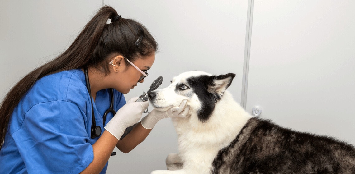
Diagnosing cherry eye in dogs is rather simple. A veterinarian will gather a general history and perform a physical examination on your dog. This will include an ophthalmic examination to evaluate the affected eye. With the history, breed, and physical examination in mind, your veterinarian will come to a diagnosis of cherry eye and then will prepare you for the next steps.
How to Treat Cherry Eye in Dogs?
Treatment of cherry eye in dogs is almost always surgical as the condition will not resolve with medical management alone. In the early phases, or before surgery can be performed, your veterinarian may prescribe an anti-inflammatory medication after ruling out any corneal ulcer formation. This medication is to alleviate any inflammation of the gland and eye while you contemplate which option is best for your dog.
There are several surgical options available for dogs with cherry eye. In the past, veterinarians have removed this gland to resolve the issue. This is a less-than-ideal technique because the gland should be preserved if at all possible. Because the gland produces a large amount of tears, partially or completely removing the gland will predispose a dog to keratoconjunctivitis sicca, which is a painful condition caused by inadequate tear production that can lead to corneal drying, ulceration, keratitis, and potential loss of vision.
Other techniques include replacing the gland or anchoring it within the lower structure of the eye. These are called envelope or pocket techniques. You may discuss these options with your family veterinarian or request a referral to a veterinary ophthalmologist.
How to Prevent Cherry Eye in Dogs?
Unfortunately, cherry eye is not a preventable disease in dogs. The underlying cause is not known at this time and, unfortunately, some dogs are just predisposed to the condition.
Before purchasing or adopting a predisposed breed, have a conversation with the breeder and ask if any other dogs in the litter or from the breeding line have been affected by this condition. If you can’t ask, be prepared that your puppy or dog may develop this condition at a young age. For this reason, it is important to purchase pet insurance to cover any mishaps that may happen in the future.
Cherry eye in dogs is caused by the prolapse of the gland within the nictitating membrane. Partial prolapses or inflammation of the gland may wax and wane, especially when treated with anti-inflammatory medication. Unfortunately, the condition will not go away on its own. Once prolapsed, the gland often stays prolapsed until surgery is performed. Ideally, the condition should be treated sooner rather than later to preserve the health of the eye.
Unfortunately, veterinarians do not know the cause of cherry eye in dogs. What we do know is that there is a genetic predisposition in affected breeds. Before purchasing a new puppy or dog, discuss the risks of hereditary conditions with the breeder.
The cost of treatment of cherry eye in dogs is dependent on the clinic, geographic location, and experience of the veterinarian performing the surgery. Surgery performed by a board-certified ophthalmologist may be more expensive but comes with specialty expertise. Generally, cherry eye surgery may range from $1,500-$4,000.

Dr. Paula Simons is an Emergency and Critical Care Veterinary Resident who aspires to be a veterinary criticalist. Dr. Simons is passionate about supporting pets and humans during their times of need. She has a special interest in critical care nutrition, trauma, and pain management. In her free time, she loves plant shopping, hiking, and traveling. She has volunteered in several different countries to help animals in need. She has two cats, Moo and Kal.


